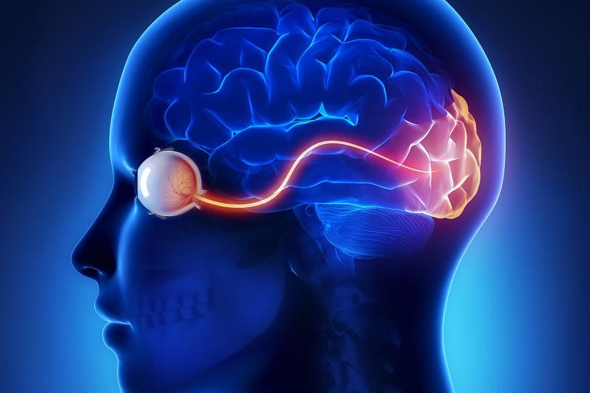
SINGAPORE – It is said that the eyes are the windows to the soul. Researchers at the Eye Research Institute of Singapore (Seri) have now discovered that they are also a peephole into brain health.
Researchers used non-invasive eye scans to link changes in the retina to what was happening in the brains of Alzheimer’s patients. Such changes can be detected early in the disease, when patients show signs of mild cognitive impairment.
By studying differences in the thickness of nerves and the density of blood vessels in the eye, researchers can now predict and detect early signs of Alzheimer’s disease, the most common type of dementia.
The eyes are organs directly connected to the brain, and the nerves and blood vessels are similar. So when one of these organs becomes diseased, the other can also undergo noticeable changes, said Seri clinician-scientist Jacqueline Chua.
“The eye is the only place in the body where you can actually visualize both neurons and blood vessels without cutting people,” she said, adding that the most standard diagnosis for Alzheimer’s disease is through brain imaging and a lumbar puncture. He added that there is.
An older study conducted at Cedars-Sinai Medical Center in the US used cadavers and tissue samples from human donors to examine potential biomarkers in the retina and correlate them with known indicators of Alzheimer’s disease. However, Seri’s team studied the retinas of living patients.
The Singapore study used devices such as optical coherence tomography (OCT) and optical coherence tomography angiography (OCTA)., which one It uses light waves to take cross-sectional pictures of a patient’s retina.
OCT allows ophthalmologists to see the characteristic layers of the retina, allowing them to map and measure retinal thickness, while OCTA takes pictures of blood vessels within and under the retina.
In the first study, Seri scientists not only looked at the thickness of the optic nerve, but also the thickness of the nerve around the macular area.
“Combining the two can actually improve detection rates much better than any single approach,” said Dr Chua, who is also an optometrist at the National Eye Center of Singapore.
In the second study, the team used OCTA to measure blood vessel density in 90 patients over the age of 50. Participants consisted of 24 participants with Alzheimer’s disease, 37 participants with mild cognitive impairment, and 29 unimpaired controls.
The researchers found that the eyes of participants with Alzheimer’s disease had significantly sparser blood vessel density compared to controls.
“These retinal imaging devices reveal neuronal thinning and microvascular changes associated with Alzheimer’s disease. With this knowledge, doctors can intervene early and suggest preventive strategies. ” said Dr. Chua.
